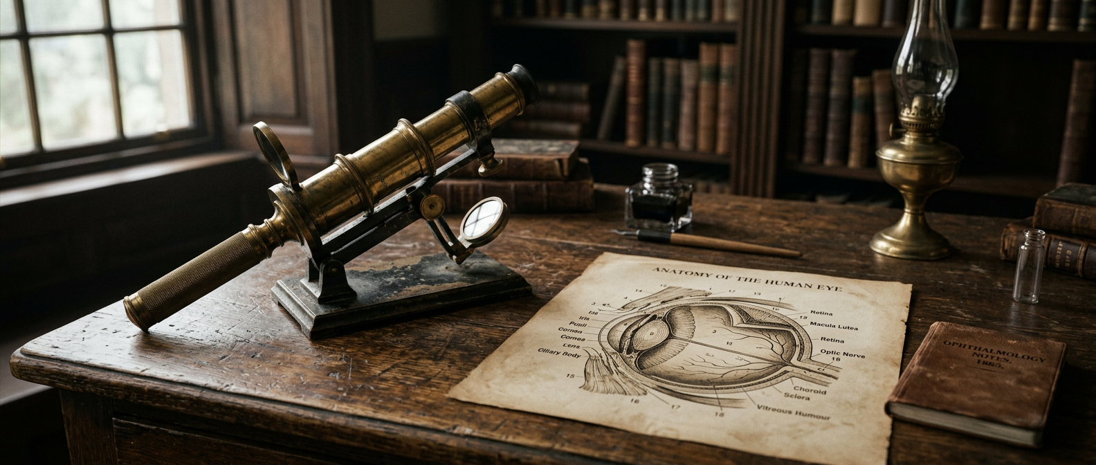
Ophthalmology · 1832 — 2026
Learning to look into the eye.
A selective international chronology with UK landmarks. Dates mark first publications, demonstrations or procedures — not the moment a technique became routine. None is a treatment for monocular vision itself; each changed how causes of sight loss are found, measured or treated.
1832
Frederick Sleigh Roberts is born.
Starting point for the Victorian case study; the later family-derived account reports childhood loss of sight in the right eye.
[1]1851
Hermann von Helmholtz publishes his ophthalmoscope; Albrecht von Graefe, Franz Cornelius Donders and William Bowman contribute to a new scientific ophthalmology.
1850s — 1880s
Ophthalmic surgery develops alongside anaesthesia, antisepsis and better instruments; cocaine is introduced as a local anaesthetic in eye surgery in 1884.
Safer examination and surgery depend on wider changes in medicine and operating practice. Moorfields records early use of cocaine in 1884.
[31]1880
William Bowman helps found the Ophthalmological Society of the United Kingdom.
Specialist institutions organise ophthalmology as a medical field; the Society is a predecessor of today’s Royal College of Ophthalmologists.
[32]1905
Eduard Zirm performs the first successful human corneal transplant.
Keratoplasty creates a surgical option for selected corneal blindness; outcomes and eligibility remain condition-specific.
[34]1911
Allvar Gullstrand demonstrates the slit lamp and receives the Nobel Prize for work on the optical system of the eye.
Slit-lamp biomicroscopy becomes foundational for examining the front of the eye; Gullstrand’s optical models inform measurement and clinical optics.
[33]1945 — 1949
Charles Schepens develops the binocular indirect ophthalmoscope; Marc Amsler introduces the Amsler grid; Harold Ridley begins intraocular lens implantation.
1950s — 1960s
Tonometry, ocular ultrasound, retinal angiography and laser photocoagulation advance; Charles Kelman introduces phacoemulsification in 1967.
Pressure measurement, imaging behind opaque media and retinal treatment expand. Phacoemulsification later transforms cataract surgery.
[32]1968 — 1970s
Trabeculectomy is developed in the UK; pars plana vitrectomy emerges; Bailey–Lovie logMAR charts arrive in 1976.
Glaucoma, vitreoretinal disease and visual function become more open to structured assessment. Acuity remains only one dimension of functional vision.
[32]1980s — 1990s
Computerised visual-field testing, excimer-laser refractive surgery and better retinal imaging develop; LASIK is introduced.
A shift toward digital measurement and elective refractive correction. Refractive surgery is not a remedy for blindness or for every cause of monocular sight.
[32]1991
David Huang, Eric Swanson, James Fujimoto and colleagues publish the first optical coherence tomography (OCT) paper.
Non-invasive cross-sectional retinal imaging, later central to diagnosing and monitoring retinal and optic-nerve disease.
[36]2000s
Intravitreal anti-VEGF medicines change the treatment of wet macular degeneration; NICE issues UK guidance in 2008.
2010s
OCT angiography, genetic diagnosis and inherited-retinal-disease research expand; the FDA approves voretigene neparvovec in 2017.
Gene therapy is a major but narrowly indicated milestone — not a general restoration of sight.
[39]2020s — 2026
Swept-source OCT, imaging analytics, tele-ophthalmology, AI-assisted screening and new gene and cell therapies develop. WHO publishes a 2026 guide to integrated vision rehabilitation.
2026 baseline: ophthalmology spans cornea, cataract, glaucoma, medical and surgical retina, paediatrics, strabismus, neuro-ophthalmology and oculoplastics. No single subspecialist covers every medical, functional and social question. [29]
Clinical interpretation
A functional state, not a single diagnosis.
A specialist assessment first seeks the cause and whether any part of it is treatable, then documents what each eye can do and how that affects everyday tasks. Acuity, refraction, fields, pupils, alignment, contrast and imaging may all be assessed — but a normal acuity result alone does not rule out field, contrast, glare, motion or binocular difficulties.
For someone relying on one eye, the health of the better-seeing eye matters most. Lenses, surgery or medication may address a remediable cause; they cannot be assumed to restore normal vision. Where useful sight cannot be restored, low-vision rehabilitation still supports activity, participation and quality of life. [40], [41]
The question is not simply “Can the person see with one eye?” It is:
- What is the diagnosis?
- What visual functions remain?
- What is changing?
- What tasks matter to the person?
- Which treatment or rehabilitation options have evidence?
- What environmental adjustments would reduce avoidable barriers?
The answer is individual, and may change over time.
Expertise
Who answers which question.
This study is an evidence synthesis until reviewed by named clinicians. An authoritative record needs specialist medicine, history, rehabilitation and lived experience together.
Ophthalmologist — neuro-ophthalmology
Is reduced vision caused by the retina, optic nerve, visual pathways or brain? How should ocular and neurological vision loss be distinguished?
Ophthalmologist — medical & surgical retina; glaucoma
Which diagnoses, prognoses, monitoring and treatments matter when a person depends on one eye or has degraded sight in it?
Ophthalmologist — cornea, cataract & ocular trauma
How do injury, corneal disease, cataract, surgery or loss of the eye change acuity, comfort, appearance and function?
Paediatric ophthalmologist & orthoptist
How do onset during visual development, amblyopia, strabismus, suppression and stereopsis affect treatment windows and later adaptation?
Optometrist & low-vision clinician
How do refraction, contrast, magnification, lighting, filters and task-specific aids affect functional vision — including without formal sight registration?
Vision rehabilitation, OT & mobility specialist
Which strategies and environmental changes help with reading, daily living, safe movement, work, education and participation?
Historian of medicine & ophthalmic archivist
What did historical terms, records, instruments and standards mean at the time? Which claims are contemporary evidence, which later retellings?
People with lived experience
Which descriptions, barriers, strategies and service gaps reflect real variation? Which assumptions should this study avoid?
An ophthalmologist can give expert medical evidence, but no single discipline can settle a person's daily experience, disability identity, access needs or rehabilitation priorities. Those require the person's own account and the work of allied professionals. [29]